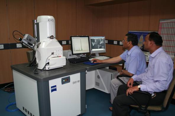
The Scanning Electron Microscope laboratory was established at WIHG in 1988 with the SEM model PHILIPS PSEM 515. This model was replaced by a new SEM with Energy Dispersive Spectrometer in 2006. The current SEM model in use is EVO 40 EP of Carl Zeiss with most advanced LN2 free EDX of Bruker make(X Flash 4010 SDD X-ray detector). The new system is fully functional with improved resolution of 3 nm and digital image processing and storage capabilities. Multiple imaging modes are available even at a pressure upto 3000 Pa using SE and BSE signals. The state of the art EDX facility is satisfying the long standing need of users, providing them qualitative and quantitative compositional analysis of different phases, grains and selected points of the sample including elemental mapping of the selected area.
The SEM-EDX is capable of capturing ultra structural details of samples as images in digital mode. The instruments can magnify specimens up to 10,00,000 times. The surface features are best seen in Secondary Electron image while compositional and topographical images can be seen with Back Scatter Electron image.
With the help of EDX, both qualitative and quantitative microanalysis can be done by nondestructive method. Elements from Boron to Uranium with a detection limit of 100 ppm can be analyzed. Besides providing routine services for mineral investigation, environmental, mineralogical, palaeontological and palaeoclimatic studies, services have been provided to outside organizations.
| SEM- Zeiss EVO-40 EP | |
| Sample viewing at normal pressure or high vacuum, with or without conductive coating (Gold or Carbon) | |
| Resolution : 30 nm ( HV SE) | |
| Magnification:upto 10,00,000 X | |
| Max. sample Dimension : Dia. - 200 mm, Height - 100 mm | |
| Sample navigation : 5 axis motorized (X, Y,Z, Tilt and rotation) | |
| Imaging mode : SE, BSE and X-ray mapping | |
| Image Store : Standard graphic file format like TIF,BMP or JPG with pixel resolution of 3072 X 2304, 16 bit |
| SDD detector with SLEW ( Super light Element Window) and Active Area- 10 mm2 | |
| Resolution: 129 eV at Mn Ka (5.98 KeV) | |
| Max. Count rate : 10,00,000 cps | |
| QUANTAX 200 - Full qualitative and quantitative analysis software with point line and area scanning and elemental mapping |
| Rocks, minerals, fossils, ores, biological, pharmaceutical, metallurgical, polymer samples, etc. are suited for SEM / EDX studies. | |
| Samples should be dry, clean and sized. | |
| Samples should be of maximum 20 mm diameter and with a height of 5mm. | |
| Samples for EDX microanalysis should be polished and made parallel to the surface of the stub. |
| Nature of samples | |
| Objective of the study | |
| Number of expected SEM micrographs to be taken | |
| Number of expected microanalysis location / spot |

